|
|
|
|
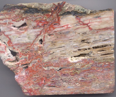
The San Quentin Sinter |
|
| A feature of
many hot springs around the world is the terrace. As water cascades down
slope from the emerging hot springs, mineral deposits build up through
time. Calcareous spring deposits are called "tufa," while siliceous spring
deposits are called "sinter." At the McLaughlin mine, the ancient terraces
that capped portions of the ore deposit were siliceous, and are appropriately
called sinter.
Because of their significance, a pictorial essay of textures found in them is presented below. All specimens are from the McLaughlin San Quentin sinter unless otherwise noted.
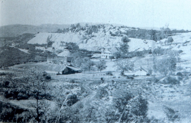
The San Quentin sinter at the McLaughlin mine was a prominent feature of the deposit. It rose above the surrounding slopes to form a low hill, and it was extensively "glory holed" in the old days to extract veins of mercury-rich cinnabar and metacinnabar ore that cut through it. The hill got its name sometime in the late Nineteenth Century, because the surface mining work was not unlike hard labor at California's first prison. In the historic photo below, San Quentin Hill appears in the background. In the foreground is the historic settlement of Johntown. Photo from Cal. State Mining Bureau Bulletin 27, 1908. |
|
 |
|
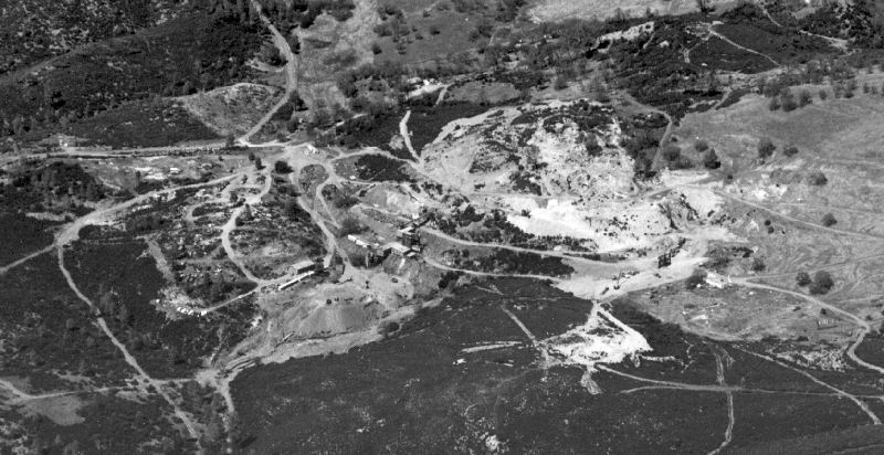
| Below, is the San Quentin Hill as it appeared from the air in 1981. The original Manhattan mercury mine furnace appears near the center of the image. Homestake Mining photo. | |
 |
|
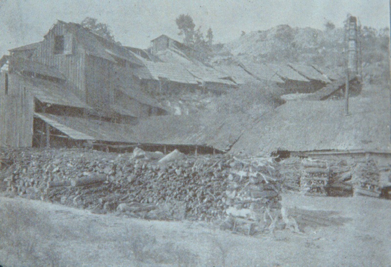
| Below, is a close-up of the old Manhattan furnace as it appeared around 1908. San Quentin Hill is on the skyline at the right of the photo. From Cal. State Mining Bureau Bulletin 27, 1908. | |
 |
|
 |
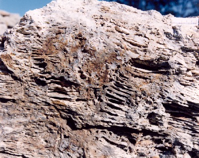
Hot springs terraces in the sinter occurred at many scales. The "terracettes" at left are in a boulder about 1 meter wide. This and many other examples of the sinter are on display in Homestake's "Stonehenge" rock display at the McLaughlin mine core library. Photo courtesy J. Farmer, NASA-Ames Research Center. |
| On a smaller scale, peculiar patterns in sinter occurred in a variety of forms. At right are ripple-like structures about 1 cm from crest to crest. D. Enderlin photo. | 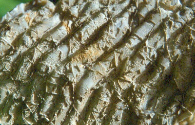 |
 |
Unlike veins, which grow from opposing walls of a cavity, sinter forms in only one direction (upward and outward from the surface on which it is depositing). Textures such as the "eggshell" layer pattern shown at left are seen in modern hot springs as well. Sinter can form as leathery mats with gas-filled cavities sometimes intervening. When lithified through time, the cavities are sometimes preserved. Many cavities are coated with crusts of quartz and chalcedony that deposited during later stages of mineralization. D. Enderlin photo. |
| Hot springs terraces can dry out as they form. As they dehydrate, they can produce mud cracks just like clayey soils do. The photo at right is a view of the top of a mud crack pattern on a sinter boulder. The cracks formed before the sinter lithified, but the cracks were completely preserved as the siliceous mud turned to stone. If you look closely at the photo of eggshell sinter above, you can see vertical structures crosscutting the eggshell layers. These are profile views of the same mudcracks. D. Enderlin photo. | 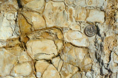 |
 |
As mud cracks developed on the sinter terraces, they occasionally curled. In extreme cases, they rolled up to form scroll-like shapes. The image at left shows such a "mud scroll" in San Quentin sinter. The structure was first thought to be a plant fragment, but closer inspection revealed its true origin. D. Enderlin photo. |
| A close-up end view of "mud scrolls" from the San Quentin sinter appears at right. D. Enderlin photo. | 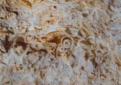 |
 |
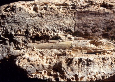
At left is another peculiar texture of the hot springs sinter at the McLaughlin mine. The spherical forms are called "geyser pearls" or "geyser eggs." Like marine oolites, these structures formed as concentric bands of precipitate built up on constantly agitated sand grains. Agitation prevented the grains from cementing to the floor of the hotspring pool as they grew. Each "pearl" contains a nucleus of rock. D. Enderlin photo. |
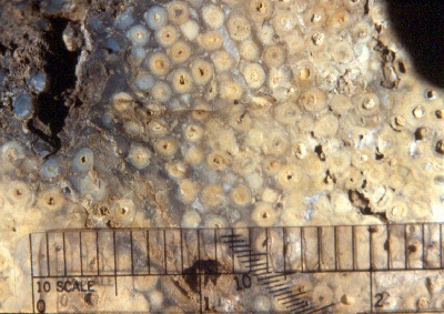
| At right is a close-up view of another "geyser pearl" specimen that reveals the internal structure. Each sphere is about 1 mm in diameter, although examples as large as a centimeter have been found at the McLaughlin deposit. In a given specimen, all grains tend to be approximately the same diameter. D. Enderlin photo. | 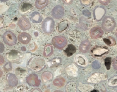 |
 |
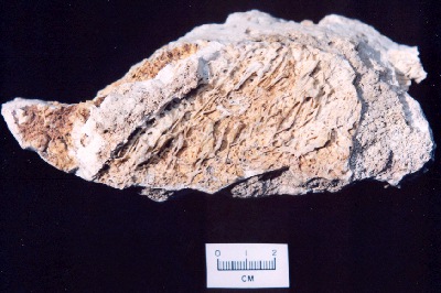
At left is a collapse breccia. Where hot springs pools were less turbulent, lily pad-like protrusions of sinter would grow outward across the water. Occasionally, these structures would collapse under their own weight or by external forces, causing them to fragment and settle to the bottom of the pool. In time, they would be cemented together by new sinter. The McLaughlin deposit contained a variety of breccias. Collapse breccia in sinter was distinguished by its clast content consisting only of sinter. In other types of breccia at McLaughlin, a variety of clast lithologies are found. |
| Sinter ranged through a variety of colors, but the most common color was white (as shown at right). In general the sinter was devoid of metal impurities, and so had no pigmenting agents. D. Enderlin photo. | 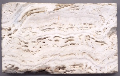 |
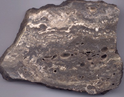 |
Dark
sinter such as that shown at left was pigmented by hydrocarbon residue.
Vein matter at depth also often displayed resiny shades of brown and tan
in response to the presence of hydrocarbons. Dark sinter typically deposited
as the first layer after a hydrothermal explosion event.
Prior to the fracturing event, fluids would vertically partition in the water column, with oils rising to the top. The "first flush" of fluids to emerge following hydrofracturing would naturally be elevated in oils. These fluids also tended to be enriched in metals. The bubble-like cavities seen in the center of this specimen are "froth veins." These structures formed when silica precipitated around globules of oil and other fluid. D. Enderlin photo. |
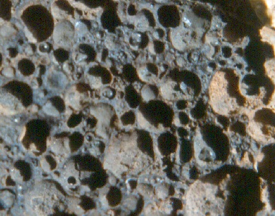 |
At left is a close-up image of another froth vein specimen from the San Quentin sinter that reveals tarry and pyrobituminous globules still in place in several of the cavities. Froth veins are not limited to sinter. Similar textures are found at many of the epithermal mercury deposits of the California Coast Ranges. D. Enderlin photo. |
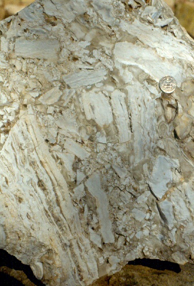
| So how does one distinguish sinter deposits from veins in a system like McLaughlin? Symmetry is the answer. Veins have bilateral growth symmetry, while sinters are unilateral. The image at right shows a vein cross-cutting sinter. Note that the banding in the sinter is convex in only one direction, which is the direction of growth. D. Enderlin photo. |  |
 |
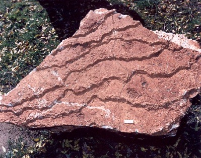
As the sinter pile grew progressively thicker, the older layers at the bottom of the pile would be cut by new generations of veins. Each generation of vein was a fracture that fed successively higher layers on the terrace. The image at left shows an opalized cinnabar vein (myrickite) crosscutting sinter. D. Enderlin photo. |
| One of the more peculiar forms observed in the San Quentin sinter was actually a fossil. The thread-like strands on the surface of the sinter sample at right are fossilized thermophyllic filamentous bacteria. The bacterial strands are completely replaced by chalcedony, but they preserve the strand-like forms from the time when they draped across the surface of a terrace, aligning with the direction of flow of the water. No other evidence of fossil life was found in the San Quentin sinter. Photo courtesy J. Farmer, NASA-Ames Research Center. | 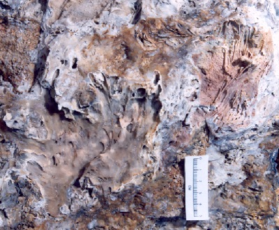 |
 |
Another example of fossilized filamentous bacteria is shown at left. Although hot springs algae can also produce such forms, the temperature of these springs was too hot to support anything but bacteria near the vents. As a well-preserved record of primitive life, the McLaughlin fossils were studied by exobiologists from the Mars Program in order to visualize what fossilized extraterrestrial primitive life might look like. Photo courtesy J. Farmer, NASA-Ames Research Center. |
| One has to give credit to the bacteria for their tenacity! Not only were fossilized remnants found at the surface, but they were also identified in vein matter over 500 feet beneath the San Quentin. The photo at right shows fossil filamentous bacteria coating a quartz vein. The sample was collected from the sheeted vein zone near the ultimate bottom of the South Pit. D. Enderlin photo. | 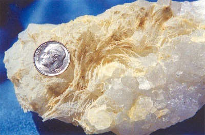 |